
Read and download free pdf of CBSE Class 12 Biology Human Reproduction Worksheet Set B. Download printable Biology Class 12 Worksheets in pdf format, CBSE Class 12 Biology Chapter 3 Human Reproduction Worksheet has been prepared as per the latest syllabus and exam pattern issued by CBSE, NCERT and KVS. Also download free pdf Biology Class 12 Assignments and practice them daily to get better marks in tests and exams for Class 12. Free chapter wise worksheets with answers have been designed by Class 12 teachers as per latest examination pattern
Chapter 3 Human Reproduction Biology Worksheet for Class 12
Class 12 Biology students should refer to the following printable worksheet in Pdf in Class 12. This test paper with questions and solutions for Class 12 Biology will be very useful for tests and exams and help you to score better marks
Class 12 Biology Chapter 3 Human Reproduction Worksheet Pdf
MULTIPLE CHOICE QUESTIONS
Question. Both corpus luteum and macula lutea are
(a) found in human ovaries
(b) a source of hormones
(c) characterized by a yellow colour
(d) contributory in maintaining pregnancy
Answer. C
Question. The early human embryo distinctly possesses
(a) gills
(b) gill slits
(c) external ear (pinna)
(d) eyebrows
Answer. C
Question. The phase of menstrual cycle in humans that lasts for 7-8 days, is
(a) follicular phase
(b) ovulatory phase
(c) luteal phase
(d) menstruation
Answer. B
Question. Which one of the following statements with regard to embryonic development in humans is correct?
(a) Cleavage divisions bring about considerable increase in the mass of protoplasm.
(b) In the second cleavage division, one of the two blastomeres usually divides a little sooner than the second.
(c) With more cleavage divisions, the resultant blastomeres become larger and larger.
(d) Cleavage division results in a hollow ball of cells called morula.
Answer. A
Question. Which one of the following events is correctly matched with the time period in a normal menstrual cycle?
(a) Release of egg : 5th day
(b) Endometrium regenerates : 5-10 days
(c) Endometrium secretes nutrients for implantation : 11-18 days
(d) Rise in progesterone level : 1-15 days
Answer. B
Question. In humans, what is the ratio of the number of gametes produced from one male primary sex cell to the number of gametes produced from one female primary sex cell?
(a) 1:3
(b) 1:4
(c) 3:1
(d) 4:1
Answer. D
Question. Corpus luteum is a mass of cells found in
(a) brain
(b) ovary
(c) pancreas
(d) spleen
Answer. B
Question. Cells of leydig are found in
(a) Testes of frog
(b) Testes of rabbit
(c) Kidney of frog
(d) Kidney of rabbit
Answer. B
Question. Meroblastic cleavage refers to which type of division of egg
(a) Complete
(b) Spiral
(c) Incomplete
(d) Horizontal
Answer. C
Question. Which of the following organ is differentiated first during development?
(a) Heart
(b) Skin
(c) Brain
(d) Neural tube
Answer. A
Question. The correct sequence of spermatogenetic stages leading to the formation of sperms in a mature human testis is:
(a) spermatogonia-spermatid-spermatocytesperms
(b) spermatocyte-spermatogonia-spermatidsperms
(c) spermatogonia-spermatocyte-spermatidsperms
(d) spermatid-spermatocyte-spermatogoniasperms
Answer. C
Question. All of the following statements concerning pregnancy are accurate EXCEPT
(a) the detection of human chorionic gonadotropin in the urine forms the basis for pregnancy tests.
(b) the cyclic release of pituitary gonadotropins and ovarian steroids is continued.
(c) the mammary gland tissue of the pregnant woman is stimulated to develop by placental hormones.
(d) the corpus luteum of pregnancy maintains the uterus until the placenta is well established.
Answer. B
Question. The following graph of relative concentrations of the four hormones present in the blood plasma of a woman during her menstrual cycle. Identify the hormones.
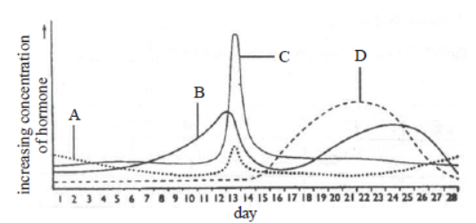
A B C D
(a) FSH Progesterone LH Oestrogen
(b) LH Progesterone FSH Oestrogen
(c) FSH Oestrogen LH Progesterone
(d) LH Oestrogen FSH Progesterone
Answer. C
Question. Ejaculation of human male contains about 200 – 300 million sperms, of which for normal fertility ____ % sperms must have normal shape and size and at least ____% must show energetic motility.
(a) 40, 60
(b) 50, 50
(c) 60, 40
(d) 30, 70
Answer. C
ASSERTION REASON QUESTIONS
Directions : These questions consist of two statements, each printed as Assertion and Reason. While answering these questions, you are required to choose any one of the following five responses.
(a) If both Assertion and Reason are correct and the Reason is a correct explanation of the Assertion.
(b) If both Assertion and Reason are correct but Reason is not a correct explanation of the Assertion.
(c) If the Assertion is correct but Reason is incorrect.
(d) If both the Assertion and Reason are incorrect.
(e) If the Assertion is incorrect but the Reason is correct.
Question. Assertion : During fertilization only head of spermatozoa enters egg.
Reason : If several spermatozoa hit the egg at same time, all can enter the egg.
Answer. C
Question. Assertion : In morula stage, cells divide without increase in size.
Reason : Zona pellucida remains undivided till cleavage is complete.
Answer. A
Question. Assertion : Death is one of the important regulatory process on earth.
Reason : It avoids over-crowding caused by continuous reproduction.
Answer. A
Question. Assertion : Old age is not an illness. It is a continuation of life with decreasing capacity for adaptation.
Reason : Cessation of mitosis is a normal genetically programmed event.
Answer. C
Question. Assertion : Senescence is the time when age associated defects are manifested.
Reason : Certain genes may be undergoing sequential switching on and off during one's life.
Answer. A
Question. Explain the role of pituitary and the ovarian hormones in menstrual cycle in human females.
Answer. Menstrual cycle is regulated by certain hormones, some of which are secreted by the pituitary gland. The pituitary gland is stimulated by releasing factors produced in hypothalamus. The hormones produced by pituitary gland influence the ovaries, which in turn affect the uterus. Anterior pituitary gland secretes two hormones FSH and LH. FSH stimulates maturation of follicle and stimulate it to secrete estrogens. Rapid secretion of LH (LH surge) induces rupturing of Graafian follicle, thereby leading to release of ovum (ovulation).
Ovary secretes two hormones : estrogen and progesterone. Estrogen stimulates follicular development and proliferation of the endometrium of the uterine wall. Progesterone produced by corpus luteum helps to maintain endometrium which is required for implantation of the fertilised ovum and other events of pregnancy.
Question. When does the corpus luteum degenerate?
Explain the immediate consequences of its degeneration in human female.
Answer. In the absence of fertilisation, the corpus luteum degenerates. Degeneration of the corpus luteum leads to decrease in the production of progesterone. As progesterone is needed for the maintenance of the endometrium, its reduction leads to disintegration of endometrium thus causing menstruation.
Question. Enumerate the events in the ovary of a human female during:
(a) Follicular phase,
(b) Luteal phase of menstrual cycle.
Answer. (a) During follicular phase, ovarian follicle secretes estrogens under the influence of FSH secreted by anterior pituitary. Estrogens stimulate the proliferation of the endometrium of the uterine wall. (b) During luteal phase, the remaining cells of the ovarian follicles after ovulation are stimulated by LH to develop corpus luteum. The corpus luteum secretes progesterone that stimulates the uterine glands to produce increased amount of mucus and is also essential for the maintenance of the endometrium which is necessary for implantation of the fertilised ovum.
Question. (a) Explain the menstrual phase in a human female. State the levels of ovarian and pituitary hormones during this phase.
Answer. (a) In a 28 day menstrual cycle, the menses takes place on days 3-5. The production of LH from the anterior lobe of the pituitary gland is considerably reduced. The withdrawal of this hormone causes degeneration of the corpus luteum and, therefore, progesterone production from the ovary is reduced. Production of estrogens from the ovary is also reduced in this phase. The endometrium of the uterus breaks down and menstruation begins. The cells of endometrium secretions, blood and the unfertilised ovum constitute the menstrual flow.
(b) During follicular phase, follicle stimulating hormone (FSH) stimulates the ovarian follicle to secrete estrogens, which in turn stimulate the proliferation of the endometrium of the uterine wall. As a result, endometrium becomes thicker by rapid cell multiplication and is accompanied by an increase of uterine gland and blood vessels. Hence, this phase is also referred as proliferative phase.
(c) At the time of ovulation, rapid secretion of LH induces rupturing of Graafian follicle, thereby releasing ovum. After ovulation has taken place, LH stimulates cells of ovarian follicle to develop corpus luteum. Corpus luteum secretes large amount of progesterone.
Question. Describe the roles of pituitary and ovarian hormones during the menstrual cycle in a human female.
Answer. Menstrual cycle in a human female consists of menstrual phase, proliferative phase (follicular phase), ovulatory phase and secretory phase (luteal phase). Days 1-5 of the cycle are known
as the menstrual phase. During this phase, menstruation occurs. At the beginning of this stage, levels of progesterone and estrogen have dropped dramatically because of the degeneration of the last cycle’s corpus luteum. This triggers the shedding of endometrium. The detached portion of the endometrium as well as blood will pass through the vagina as the menstrual flow. Days 5-14 are known as the proliferative phase. Proliferative phase consists of growth of endometrium of uterus, Fallopian tube and vagina. In ovary, a Graafian follicle grows, matures and secretes estrogen during this phase. The endometrium grows thicker and becomes more vascularised and glandular. Change in the levels of pituitary and ovarian hormones bring about these changes in the ovary and uterus. The levels of LH and FSH increase gradually during the follicular phase and stimulate follicular development as well as secretion of estrogens by the growing follicles
In ovulatory phase, both LH and FSH attain a peak level in the middle of cycle (about 14 day). Rapid secretion of LH leading to its maximum level during
the mid-cycle called LH surge induces rupture of Graafian follicle and thereby the release of ovum (ovulation).
After ovulation and in response to luteinising hormone, the portion of the Graafian follicle that remains in the ovary enlarges and is transformed into a corpus luteum containing yellow substance (called lutein) and the luteal phase begins. The corpus luteum secretes large amounts of progesterone which is essential for maintenance of endometrium. Such an endometrium is necessary for implantation of the fertilised ovum and other events of pregnancy. In the absence of fertilisation, the corpus luteum degenerates. This causes disintegration of the endometrium leading to menstruation, marking a new cycle.
Question. When and where are primary oocytes formed in human female? Trace the development of these oocytes till ovulation (in menstrual cycle). How do gonadotropins influence this developmental process?
Answer. . Primary oocytes are formed inside the ovary during the embryonic developmental stage. Oogenesis is initiated during the embryonic development stage when oogonia are formed within each fetal ovary. These cells start division and enter into prophase-I of the meiotic division and get temporarily arrested at that stage, called primary oocytes. Each primary oocyte then gets surrounded by a layer of granulosa cells and is called the primary follicle. The primary follicles get surrounded by more layers of granulosa cells and a new theca and are called secondary follicles.
The secondary follicle soon transforms into a tertiary follicle which is characterised by fluid filled cavity called antrum. The theca layer is organised into an inner theca interna and an outer theca externa. The primary oocyte within the tertiary follicle grows in size and completes its first meiotic division. It is an unequal division resulting in the formation of a large haploid secondary oocyte and a tiny first polar body. The tertiary follicle further changes into the mature follicle or Graafian follicle. The secondary oocyte forms a new membrane called zona pellucida surrounding it. The Graafian follicle now ruptures to release the secondary oocyte (ovum) from the ovary by the process called ovulation.
Effect of gonadotropins : Gonadotropin releasing hormone (GnRH) is secreted by the hypothalamus, which stimulates the release of follicle stimulating hormone (FSH) and luteinising hormone (LH). FSH stimulates the ovarian follicles to produce estrogens, which stimulate the proliferation of the endometrium of uterine wall. LH causes ovulation and stimulates the corpus luteum of the ovary to secrete progesterone.
Question. Mention the function of zona pellucida.
Answer. During fertilisation, a sperm comes in contact with the zona pellucida layer of ovum which induces changes in the membrane that block the entry of additional sperms. So, zona pellucida ensures that only one sperm can fertilise an ovum.
Question. How does the sperm penetrate through the zona pellucida in human ovum?
Answer. The sperm penetrate through the zona pellucida by releasing sperm lysins.
Question. How is the entry of only one sperm and not many ensured into an ovum during fertilisation in humans?
Answer. Depolarisation of the egg plasma membrane by binding of sperm to it checks additional sperms from entering into it.
Question. Mention the function of trophoblast in human embryo.
Answer. Trophoblast helps to provide nutrition to the embryo.
Question. Name the embryonic stage that gets implanted in the uterine wall of human female.
Answer. Blastocyst
Question. Where does fertilisation occur in humans?
Explain the events that occur during this process.
Answer. The motile sperms swim rapidly, pass through the cervix, enter into the uterus and finally reach the junction of the isthmus and ampulla (ampullary- isthmic junction) of the Fallopian tube. The ovum released by the ovary is also transported to the ampullary-isthmic junction where fertilisation takes place. During fertilisation, a sperm comes in contact with the zona pellucida layer of the ovum and induces changes in the membrane that block the entry of additional sperms. The secretions of the acrosome help the sperm enter into the cytoplasm of the ovum through the zona pellucida and the plasma membrane. This induces the completion of the meiotic division of the secondary oocyte. The second meiotic division is also unequal and results in the formation of a second polar body and haploid ovum (ootid). Soon the haploid nucleus of the sperms and that of the ovum fuse together to form a diploid zygote.
Question. List the different parts of the human oviduct through which the ovum travels till it meets the sperm for fertilisation.
Answer. . Fimbriae, infundibulum, ampulla and isthmus are the main parts of oviduct, through which ovum travels till it meets the sperm for fertilisation. Finally it reach the ampullary-isthmic junction of oviduct.
Question. Mention the number of cells in the following stages.
| Embryonic stage | No. of cells |
| Zygote | (a) |
| Morula | (b) |
| Blastocyst | (c) |
Answer. (a) 1 (b) 16 (c) 64
Question. Name the embryonic stage that gets implanted in human female. Explain the events that occur during this process.
Answer. Blastocyst gets implanted in human female. In a blastocyst, the blastomeres are arranged into an outer layer called trophoblast and an inner group of cells called the inner cell mass. The trophoblast then gets attached to the endometrium and the inner cell mass gets differentiated as the embryo. After attachment the uterine cells divide rapidly and cover the blastocyst. As a result, the blastocyst becomes embedded in the endometrium of the uterus. This whole phenomenon is called implantation and it leads to pregnancy.
Question. Where is morula formed in humans? Explain the process of its development from zygote.
Answer. a) Blastocyst is shown in the figure. ‘A’ is inner cell mass and ‘B’ is trophoblast.
(b) Inner cell mass form the foetus.
Question. Briefly explain the events of fertilisation and implantation in an adult human female.
Answer. The events of fertilisation in human female are: (i) Acrosomal reaction : After ovulation, the secondary oocyte reaches the Fallopian tube. The capacitated sperm releases hydrolytic enzymes (sperm lysins) present in the acrosome, when it comes in contact with surface of egg covering. Important sperm lysins are (i) hyaluronidase that acts on the ground substances of follicle cells, (ii) corona penetrating enzyme that dissolves corona radiata and (iii) zona lysine or acrosin that helps to digest the zona pellucida. Due to acrosomal reaction, plasma membrane of sperm fuses with that of secondary oocyte and depolarisation of oocyte membrane occurs.
(ii) Cortical reaction : Immediately after the fusion of sperm and egg plasma membranes, the egg shows a cortical reaction to further check the entry of more sperms. In this reaction, the cortical granules present beneath the ovum’s plasma membrane fuse with the same and release their contents (enzymes) between it and zona pellucida. These enzymes harden the zona pellucida, which now functions as the sure block to polyspermy.
(iii) Sperm entry : The egg extends around the entering sperm, finger-like processes, called microvilli, which constitute a fertilisation cone. The latter take the entire sperm into the egg. The distal centriole of the sperm divides and forms two centrioles to generate the mitotic spindle for cell division.
(iv) Karyogamy : The sperm entry stimulates the egg (secondary oocyte) to resume and complete the suspended meiosis - II. This produces a haploid mature ovum and a second polar body. The head of sperm separates from the middle piece and tail to become male pronucleus and nucleus of ovum is called female pronucleus. The second polar body and sperm tail degenerate. Mixing up of the chromosomes of a spermatozoon and an ovum is called karyogamy or amphimixis. This completes the act of fertilisation. The fertilised ovum is now a diploid cell having 23 pairs of chromosomes, and is termed zygote.
The events of implantation are discussed as follows: Implantation is the attachment of blastocyst to the uterine wall. It occurs after 7 days of fertilisation. As zygote moves towards the uterus, it undergoes series of mitotic divisions known as cleavage and forms 2,4,8,16 daughter cells called blastomeres. The embryo with 8 blastomeres is called morula. The morula transforms into blastocyst. In a blastocyst, the blastomeres are arranged into an outer layer called trophoblast and an inner group of cells called the inner cell mass. The trophoblast then gets attached to the endometrium and the inner cell mass gets differentiated as the embryo. After attachment the uterine cells divide rapidly and cover the blastocyst. As a result, the blastocyst becomes embedded in the endometrium of the uterus. This whole phenomenon is called implantation and it leads to pregnancy
Question. Mention the site of fertilisation of a human ovum. List the events that follow in sequence until the implantation of the blastocyst.
Answer. Fertilisation occurs in ampullary-isthmic junction of Fallopian tube.
Question. Comment on the role of placenta as an endocrine gland.
Answer. The placenta acts as an endocrine gland and secretes the following hormones :
(i) Human chorionic gonadotropin (hCG)
(ii) Human chorionic somatomammotropin (hCS)
(iii) Progesterone (iv) Estrogen (v) Relaxin
(vi) Chorionic thyrotropin and (vii) Chorionic corticotropin
The hCG stimulates and maintains the corpus luteum to secrete progesterone until the end of pregnancy. The hCS stimulates the growth of the mammary glands during pregnancy. Relaxin facilitates parturition (act of birth) by soffening the connective tissues of the pubic symphysis. The level of hormones like estrogen, progesterone etc. are increased in maternal blood during pregnancy. Increased production of these hormones is necessary for supporting the fetal growth, metabolic changes in mother and maintenance of pregnancy.
Question. When and where do chorionic villi appear in humans? State their function.
Answer. After implantation, finger-like projections appear on the trophoblast called chorionic villi which are surrounded by the uterine tissue and maternal blood. The chorionic villi and uterine tissue become interdigitated with each other and jointly form a structural and functional unit between developing embryo (foetus) and maternal body called placenta, which facilitates the supply of oxygen and nutrients to the embryo and also removal of carbon dioxide and excretory waste materials produced by the embryo. Placenta also acts as an endocrine tissue and produces several hormones essential for supporting the foetal growth, metabolic changes in the mother and maintenance of pregnancy.
Question. Explain the function of umbilical cord.
Answer. The placenta is connected to embryo through which helps in the transport of substances to and from the embryo.
Question. Explain the fate of inner cell mass in a human embryo immediately after implantation.
Answer. Immediately after implantation, the inner cell mass (embryo) differentiates into an outer layer called ectoderm and an inner layer called endoderm. A mesoderm soon appears between the ectoderm and the endoderm. These three layers give rise to all tissues (organs) in adults.
Question. Write the function of oxytocin.
Answer. Oxytocin acts on the uterine muscle causing their contractions that help in the expulsion of the baby out of the uterus through the birth canal.
Question. What stimulates pituitary to release the hormone responsible for parturition? Name the hormone.
Answer. The signals from the fully developed foetus and placenta triggers release of oxytocin from the maternal pituitary. Oxytocin is responsible for parturition.
Question. Why is oxytocin called as ‘birth hormone’ ?
Answer. Oxytocin is called as ‘birth hormone’ as it causes increased contraction of the uterine muscles and thus explusion of the baby from the uterus through the birth canal.
Question. (a) Where do the signals for parturition originate from in humans?
(b) Why is it important to feed the newborn babies on colostrum?
Answer. (a)The signals for parturition originate from the fully developed foetus and the placenta which induce mild uterine contractions called foetal ejection reflex.
(b) After birth, the breast first release milk is called colostrum for 2 or 3 days. This is a thin, yellowish, fluid, often called foremilk. It contains cells from the alveoli and is rich in protein (lactalbumin & lactoprotein), antibodies, but low in fat. Breast-feeding during the initial period of infant growth is recommended by doctors for bringing up a healthy baby
Question. Why is parturition called a neuroendocrine mechanism?
Answer. Process of parturition is induced by both neural system and endocrine system therefore, it is called a neuro-endocrine mechanism. The signals for parturition originate from the fully developed foetus and the placenta which induce mild uterine contractions called foetal ejection reflex. This triggers release of oxytocin from the maternal pituitary. Oxytocin acts on the uterine muscle and causes stronger uterine contractions that leads to expulsion of the baby out of the uterus through the birth canal.
Question.How is the milk production regulated by hormones in human female? Explain.
Answer. Secretion and storage of milk generally begins after birth of young one, usually within 24 hours under the influence of hormone prolactin (PRL) secreted by anterior lobe of the pituitary gland. However, the ejection of milk is stimulated by the hormone oxytocin (OT) released from the posterior lobe of the pituitary gland.
Question. Describe the process of parturition in humans.
Answer. The act of expelling the full term young one from the mother’s uterus at the end of gestation period is called parturition. Process of parturition is induced by both nervous system and hormones secreted by the endocrine glands of the mother. The signals for child birth (parturition) originate from the fully matured foetus and placenta which induce mild uterine contractions called foetal ejection reflex. This causes quick release of oxytocin from the maternal pituitary gland.
Oxytocin acts on the uterine muscle and causes stronger uterine contractions which in turn further stimulates the secretion of oxytocin. The stimulatory reflex between the uterine contraction and oxytocin secretion continues resulting in stronger and stronger contractions. This leads to expulsion of the baby from the uterus through the birth canal.
Please click on below link to download CBSE Class 12 Biology Human Reproduction Worksheet Set B
| CBSE Class 12 Biology Evolution Question Bank |
| CBSE Class 12 Biology Evolution Worksheet Set A |
| CBSE Class 12 Biology Evolution Worksheet Set B |
| CBSE Class 12 Biology Biotechnology And Its Application Question Bank |
| CBSE Class 12 Biology Biotechnology and Its Applications Worksheet Set A |
| CBSE Class 12 Biology Ecosystem Question Bank |
| CBSE Class 12 Biology Ecosystem Worksheet Set A |
| CBSE Class 12 Biology Ecosystem Worksheet Set B |
More Study Material
CBSE Class 12 Biology Chapter 3 Human Reproduction Worksheet
The above practice worksheet for Chapter 3 Human Reproduction has been designed as per the current syllabus for Class 12 Biology released by CBSE. Students studying in Class 12 can easily download in Pdf format and practice the questions and answers given in the above practice worksheet for Class 12 Biology on a daily basis. All the latest practice worksheets with solutions have been developed for Biology by referring to the most important and regularly asked topics that the students should learn and practice to get better scores in their examinations. Studiestoday is the best portal for Printable Worksheets for Class 12 Biology students to get all the latest study material free of cost.
Worksheet for Biology CBSE Class 12 Chapter 3 Human Reproduction
Teachers of studiestoday have referred to the NCERT book for Class 12 Biology to develop the Biology Class 12 worksheet. If you download the practice worksheet for the above chapter daily, you will get better scores in Class 12 exams this year as you will have stronger concepts. Daily questions practice of Biology printable worksheet and its study material will help students to have a stronger understanding of all concepts and also make them experts on all scoring topics. You can easily download and save all revision Worksheets for Class 12 Biology also from www.studiestoday.com without paying anything in Pdf format. After solving the questions given in the practice sheet which have been developed as per the latest course books also refer to the NCERT solutions for Class 12 Biology designed by our teachers
Chapter 3 Human Reproduction worksheet Biology CBSE Class 12
All practice paper sheet given above for Class 12 Biology have been made as per the latest syllabus and books issued for the current academic year. The students of Class 12 can be assured that the answers have been also provided by our teachers for all test paper of Biology so that you are able to solve the problems and then compare your answers with the solutions provided by us. We have also provided a lot of MCQ questions for Class 12 Biology in the worksheet so that you can solve questions relating to all topics given in each chapter. All study material for Class 12 Biology students have been given on studiestoday.
Chapter 3 Human Reproduction CBSE Class 12 Biology Worksheet
Regular printable worksheet practice helps to gain more practice in solving questions to obtain a more comprehensive understanding of Chapter 3 Human Reproduction concepts. Practice worksheets play an important role in developing an understanding of Chapter 3 Human Reproduction in CBSE Class 12. Students can download and save or print all the printable worksheets, assignments, and practice sheets of the above chapter in Class 12 Biology in Pdf format from studiestoday. You can print or read them online on your computer or mobile or any other device. After solving these you should also refer to Class 12 Biology MCQ Test for the same chapter.
Worksheet for CBSE Biology Class 12 Chapter 3 Human Reproduction
CBSE Class 12 Biology best textbooks have been used for writing the problems given in the above worksheet. If you have tests coming up then you should revise all concepts relating to Chapter 3 Human Reproduction and then take out a print of the above practice sheet and attempt all problems. We have also provided a lot of other Worksheets for Class 12 Biology which you can use to further make yourself better in Biology
You can download the CBSE Practice worksheets for Class 12 Biology Chapter 3 Human Reproduction for the latest session from StudiesToday.com
Yes, you can click on the links above and download chapter-wise Practice worksheets in PDFs for Class 12 for Biology Chapter 3 Human Reproduction
Yes, the Practice worksheets issued for Chapter 3 Human Reproduction Class 12 Biology have been made available here for the latest academic session
You can easily access the links above and download the Class 12 Practice worksheets Biology for Chapter 3 Human Reproduction
There is no charge for the Practice worksheets for Class 12 CBSE Biology Chapter 3 Human Reproduction you can download everything free
Regular revision of practice worksheets given on studiestoday for Class 12 subject Biology Chapter 3 Human Reproduction can help you to score better marks in exams
Yes, studiestoday.com provides all the latest Class 12 Biology Chapter 3 Human Reproduction test practice sheets with answers based on the latest books for the current academic session
Yes, studiestoday provides worksheets in Pdf for Chapter 3 Human Reproduction Class 12 Biology in mobile-friendly format and can be accessed on smartphones and tablets.
Yes, practice worksheets for Class 12 Biology Chapter 3 Human Reproduction are available in multiple languages, including English, Hindi